Craniomaxillofacial Surgery
Overview
The specialty of craniofacial surgery involves the treatment of skull and facial deformities arising at the time of birth. The Department of Craniomaxillofacial Surgery at Amrita Institute of Medical Sciences with its state of art facilities and outstanding team of doctors offers the highest quality of care. The philosophy of care for the child with a craniofacial condition is to provide a comprehensive multidisciplinary service that addresses both the equally important functional, aesthetic and psychological aspects of the conditions seen by the group. The multidisciplinary approach includes a Craniofacial Surgeon, Paediatric Neurosurgeon, Neuro-Anesthetist, Paediatrician, Ophthalmologists, Plastic Surgeons, Audiologists and Dentists.
Facilities Available
- Naso-alveolar Molding (NAM)
- The Lip Repair (Cheiloplasty)
- The Palate Repair (Palatoplasty)
- Surgery for the Gum (Secondary alveolar bone grafting)
- Surgery for Speech (Pharyngoplasty)
- Surgery for Hearing
- Orthodontic Treatment
- Jaw Surgery (Osteotomy/ Distraction Osteogenesis)
- The Nose Repair (Cleft Rhinoplasty)
Services Offered
Cleft lip and palate deformities are managed by a multi-disciplinary team involving intensive care specialists, Cleft surgeon, Orthodontist, speech and language pathologist and clinical psychologist, with the care starting from birth till completion of growth of the child.
Cleft lip and cleft palate are splits or gap in the upper lip, the roof of the mouth (palate) or both. Cleft lip and cleft palate result when facial structures do not fuse inside the womb.
Cleft lip and cleft palate are among the most common birth defects nearly 1 in every 800 – 1000 live births. They most commonly occur as isolated birth defects but are also associated with many inherited genetic conditions or syndromes. Cleft lip and palate can occur alone or in combination with each other.
The issues associated with cleft lip and cleft palate can be corrected by a series of surgeries and supportive therapy which not only restore normal function but gives near normal appearance with minimal scars.
The immediate concern following birth of the child is with regard to adequate feeding, which is taught to the mother. Positioning the child Upright while feeding and use of special feeding nipples will help children with cleft palate to feed well.
Treatments
The treatments offered at our department are:
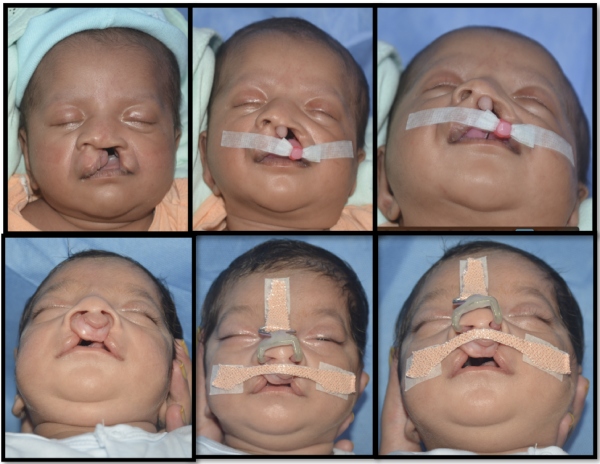
Nasal and Alveolar Molding (NAM)
Wide clefts deform the nose and gums of upper jaw and the inability of the child to create suction in the mouth due to defect in the upper jaw makes feeding difficult for the child. An oral appliance can be used (preferably given within 1-3 weeks after birth) to help with feeding the child and tp reshape the deformed gums and nasal cartilages as a preparation for the cleft lip surgery in order to achieve a more optimal outcome.
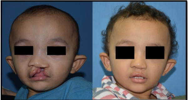
Cleft Lip Surgery
Cleft lip/ nose surgery (cheilo-rhinoplasty) is undertaken after completion of NAM therapy and is performed usually after 3 months of age, once child achieves 5 Kg of weight.
Surgery is performed in the hospital under general anesthesia. The child is admitted to the hospital a day prior to the procedure and the surgery requires an average of 2 hours. The hospital stay is usually 48 hours post-operative.
Stitches on the lip are removed 7 - 10 days after surgery. Specific instructions regarding this procedure will be given to the parents at discharge.
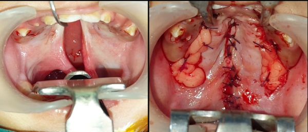
Cleft Palate Surgery
Clefts of the palate can occur alone, as an isolated deformity, or in combination with a unilateral or bilateral cleft of the lip. In twenty-five to thirty percent of all individuals with a cleft deformity, the cleft palate is the only cleft problem.
Cleft palate surgery is crucial for feeding, development of speech and to avoid middle ear infections. The surgery takes approximately 2 hours.Tissues from both sides are brought together to the center to recreate a complete palate.
Interim Treatment
Speech Therapy
For children with cleft palate Speech therapy is initiated after the age of 2 years for development of good speech.
Primarily for social reasons, minor preschool correction procedures are undertaken for lip or nose after the age of 5-6 years, when needed.
Whenever indicated, secondary surgical procedures for lengthening the palate can be undertaken for children who do not attain good speech with speech therapy alone, after the age of 6. Assessment by speech therapist as well as investigations like video fluoroscopy and nasal endoscopy are used to evaluate the quality and improvement in speech before and after surgery.

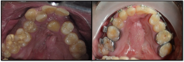
Orthodontic Treatment (Braces)
The orthodontist is an integral caregiver for the cleft child, who treats the child throughout his/her growth period. After age of 8 years, orthodontist will monitor the child on a routine basis to assess need for jaw expansion to prepare the child for alveolar bone grafting surgery.
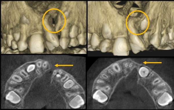
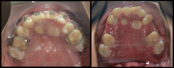
Cleft Alveolus Surgery
An alveolar bone graft performed at the age of 8 -10 years, to add bone to the gum ridge in a child who was born with both a complete cleft lip, cleft palate and cleft alveolus. Bone is harvested from hip bone and used to fill the bony gap in the gums.
The alveolar bone graft is done when canine tooth completes three-fourth of its development inside the bone and is monitored with help of periodic X-rays. This surgery is important for the proper eruption of permanent teeth of the child, after the milk teeth are shed.
The surgery takes about 2 hours and has no long-term negative effect on walking and growth of the child. The child is asked to avoid strenuous activities for 4-6 weeks. The child can resume normal activities once healing is complete.
Once the canine tooth erupts in to mouth after bone grafting the orthodontist will take measures to guide the tooth into a proper position in the arch. Face-mask therapy may be started at this age to bring the upper jaw forward to address its reduced development when compared to lower jaw.
Anterior Maxillary Distraction
Reduced size and growth of the upper jaw cause crowding of teeth, lead to poor speech and facial deformity during the pre-pubertal growth phase of the child. Whenever orthodontic expansion does not suffice, surgically assisted lengthening and widening of the upper jaw can be performed to gain adequate length and width of the upper jaw which will help the orthodontist to relieve the crowding and align the teeth. The increased space for tongue created by procedure makes articulation of speech better and at the same time improves facial appearance which helps with better social integration of the child with his/her peer group.
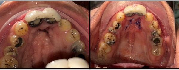
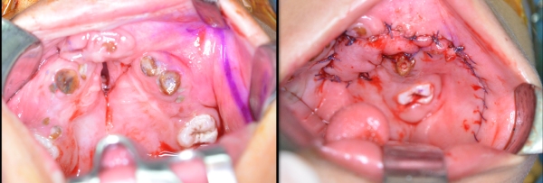
Palatal Fistula Closure
At times, wound breakdown after cleft palate surgery can lead to communication between mouth and nose which are called fistulae. Presence of fistula can cause nasal regurgitation of solid or liquid food and nasal tone of voice during speech. In young children persistent fistula present in the gum region should be closed to prepare for bone grating and orthodontic treatment. Fistula closure can be done using different methods like a simple mobilization of marginal tissue, turn over flap from surrounding tissue, or bringing in adjacent tissue from cheek (buccal flap / labial flap / FAMM flap) or from the tongue (tongue flap). A successful closure of fistula can alleviate the discomfort from nasal regurgitation of food particles and improve the voice quality by reducing nasality of speech.
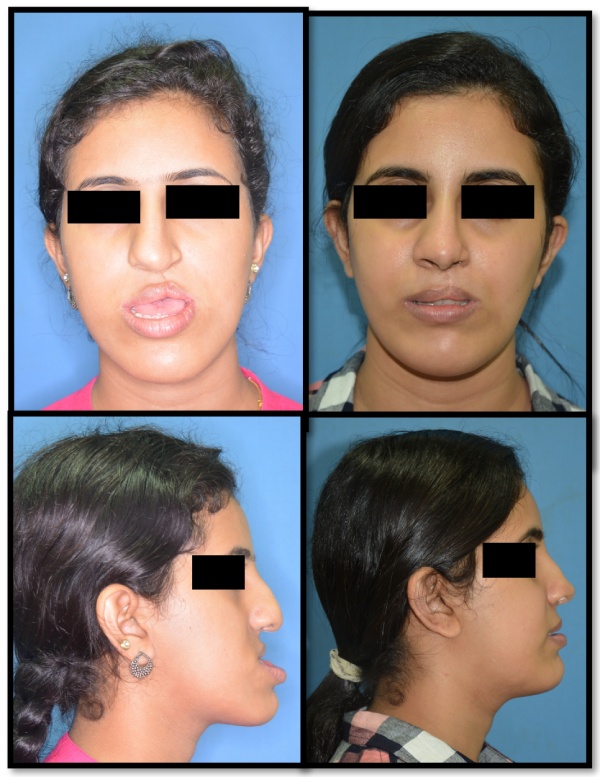
Orthognathic Surgery
Patients are kept under follow up of the doctors throughout the phase of growth. If upper jaw growth deficiency becomes further evident during post pubertal growth spurt, the child will be planned for jaw (Orthognathic) surgery. Even though the procedure is done after completion of growth (15-16 for girls, 18 for boys), orthodontic preparation is started much earlier. Orthognathic surgery brings the jaw forward to improve the bite and facial appearance. Post operative healing requires about 2 months and hence treatment is planned to coincide the surgery with academic break between school and college admissions.
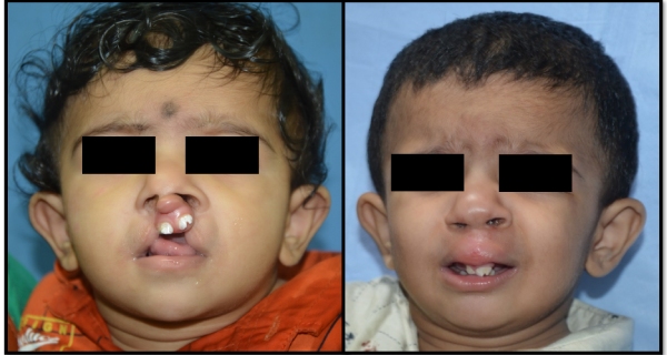
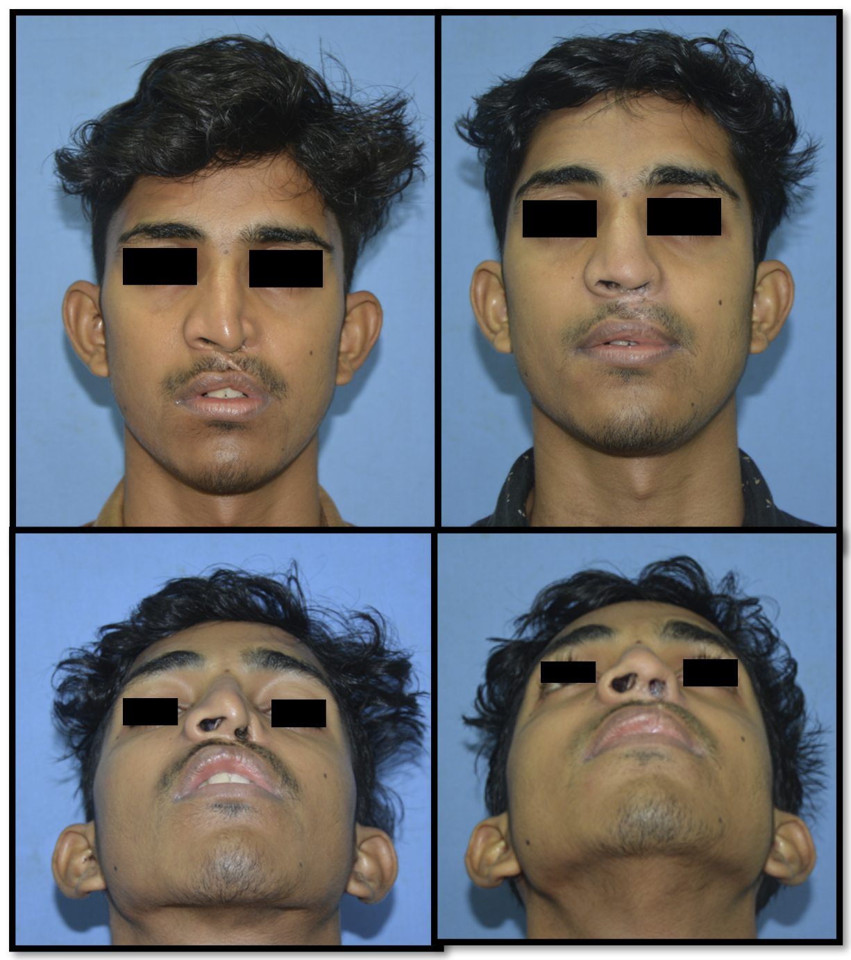
Cleft Rhinoplasty (Nose Surgery)
Cleft Rhinoplasty is the reshaping of the nose of cleft patents and is done as the final soft tissue correction after completion of all bony related procedures which create the base for nosei. Residual lip deformities if any are also corrected at this time. The surgery takes approximately 3 hours. The hospital stay is mostly for 2 days and patient can resume the normal activities after 15 - 20 days of Surgery.
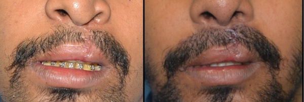
Mustache Transplant
Hair follicles from beard are grafted to the cleft lip scar in male patients to provide a full mustache.
Pierre Robin Sequence
- Airway management
- Cleft palate surgery
- Speech therapy
- Orthodontics
For Appointments Contact: 04842851401
Craniosynostosis is the early fusion of the cranial suture / sutures leading to distorted shape of the head. Surgical correction of craniosynostosis involves remodeling / reshaping of the skull and not the brain. The ideal time for correction of craniosynostosis is before one year of age. As age increases the deformity increases.
- Plagiocephaly (Unicoronal Synostosis): It is the commonest craniofacial condition caused due to premature / early fusion of the coronal suture on one side (one half of the coronal suture) characterized by an asymmetrical distortion of the skull on the affected side (flattening of the forehead on the affected side and increased forehead bulge on the opposite side.
- Brachycephaly (Coronal Suture Synostosis): Caused due to the premature fusion of the entire coronal suture. Children with brachycephaly often have an increased height of the forehead and flat forehead.
- Trigonocephaly (Metopic Suture Synostosis): Head appears to be triangular in shape.
- Scaphocepahly (Sagittal Suture Synostosis): Bullet shaped head, often presents with an increased head length.
- Multi-sutural Synostosis: There is fusion of more than cranial suture, which leads to a complete deformation of the head.
Facial cleft is an opening or gap in the face, or a malformation of a part of the face. All structures like bone, soft tissue, skin etc. can be affected. Facial clefts are extremely rare congenital anomalies.
Hypertelorism is an abnormally increased distance between the orbits (eyes). Usually associated with facial cleft.
- Aperts syndrome
- Crouzons syndrome
- Saethre-Chotzen syndrome
- Pfeiffers syndrome
The issues associated with these conditions are increased intracranial pressure, compromised airway, visual disturbances (vision), sleep apnea, altered head shape. To correct the increased intra cranial pressure and to improvise the vision, posterior cranial vault is considered as an initial step. Once the intracranial pressure is relieved, the midface advancement and cranioplasty is done to improve the airway (breathing ability) and appearance (correction of head shape).
ROSA Robotic Surgical Assistant is used to better precision in carrying out the surgical procedure.
- Cranio-fronto-nasal dysplasia
- Encephalocele
- Hemifacial microsomia.
- Treacher-Collins
- Goldenhar/hemifacial microsomia.
Video Gallery
Contact Us
Phone: 0484 – 2851401, 0484 6681401
Email: [email protected]
Doctors