Fetal Cardiology Division
1 DoctorsOverview
The fetal cardiology division at Amrita Hospital, Kochi was launched in January 2008. The aim of this service is to create awareness among the medical community regarding the need and potential benefits of prenatal diagnosis of birth defects of the heart. This will provide the concerned families with more options for management rather than facing the trauma of taking a decision after the baby is born. The concept of prenatal diagnosis and planned peri-partum care for fetuses with critical heart defects was pioneered by us in India and South Asia. The division is very active academically and has been instrumental in the training of healthcare professionals in fetal heart evaluation globally and offered training courses and fellowships in fetal cardiology for doctors (both in-person and online). The division has published several important research papers in the field of Fetal Cardiology in various peer reviewed national and international journals. The division is spearheaded by Prof. Balu Vaidyanathan, who is renowned globally for his contributions to the field of Fetal Cardiology.
Doctors

Why choose us?
- Pioneered prenatal diagnosis and planned perinatal care for fetuses with critical heart defects in India.
- Provides delivery services and expedited neonatal cardiac care under one roof.
- Established in 2008, with over 10,000 fetal echocardiography studies conducted so far.
- Enabled planned delivery of more than 750 women carrying fetuses with critical heart defects at Amrita Hospital, Kochi.
- Performed over 500 neonatal cardiac procedures with a success rate of over 98%.
- Treated fetal heart rhythm disorders in 100+ fetuses through in-utero therapy.
- Performed the first successful in-utero fetal aortic valve dilatation in 2015.
What We Offer
- Comprehensive care and treatment for various heart problems in the developing fetus.
- State-of-the-art diagnostic equipment, Voluson Expert 22 and 4D STIC Ultrasound, for early detection of fetal heart issues.
- A multidisciplinary team of specialists, including fetal cardiologists, fetal medicine experts, and fetal genetics specialists, makes the treatment decisions.
- Counselors to educate and help parents in the decision-making process
- Service coordinators to ensure seamless care for a fetus with a heart defect
- Close collaboration with allied departments, including obstetrics, radiology, pediatric cardiology, pediatric cardiac surgery, and neonatology, for better outcomes
Service
- Comprehensive fetal cardiac imaging and diagnosis
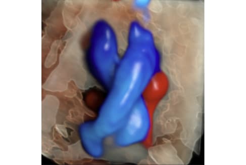
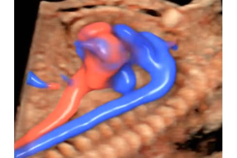
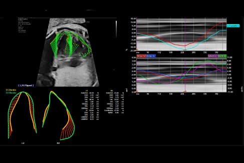
- 4D STIC Fetal echocardiography and Fetal Quantification ( fetal HQ analysis)
Facilities
- A dedicated multidisciplinary team of experts to provide flawless care for a fetus with heart disorders
- Expert management and support during pregnancy, birth, neonatal, and extended post-natal care
- Most advanced technology- including Voluson Expert 22 Ultrasound equipment with its full array of transducers and fetal HQ software- for fetal heart function assessment.
- Accurate diagnosis of fetal heart defects through real-time and offline analysis of fetal heart function evaluations.
- Parental counseling to support informed decision-making.
- Highly specialized and expedited cardiac care for babies born in our center following a prenatal diagnosis.
Fellowship Programmes/Trainings
Gallery
FAQs
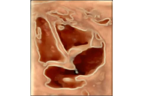
Fetal echocardiography is a highly specialized form of antenatal scan where a detailed evaluation of the unborn baby’s heart is carried out. This enables diagnosis of various forms of birth defects of the heart (congenital heart defects) and heart beat disorders (rhythm problems) in the fetus.
The principle of ultrasound scan is used for this technique. A minimum requirement for screening the fetal heart is a combination of 4 chamber view (heart chambers, valves and partitions), outflow tract view and 3-vessel view (blood vessels) which enables most of the cardiac defects to be detected. A detailed fetal echocardiography requires a thorough knowledge about the various types of birth defects of the heart and what all treatment can be offered including the costs involved. Few centers in India have a specialized fetal cardiology unit that provides a comprehensive facility for diagnosis, counseling and then treatment for various birth defects of the heart.
The ideal timing of fetal heart scan would be around 18-20 weeks. It is preferable to conduct these scans sufficiently early in pregnancy, so that if a problem is diagnosed, the family can be offered all possible options for management. Table 1 summarizes indications for fetal heart scan.
Fetal echocardiography uses the common ultrasound principle and it has been shown without any doubt that this is very safe, even if performed multiple times, in pregnancy.
Pre-natal diagnosis offers a much wider options to the expectant family, especially in the setting of developing countries with limited resources to treat complex heart defects.
Early diagnosis of very complex cardiac defects and multi-system disorders (cardiac defects with associated anomalies like genetic disorders) offers families the option of medical termination of pregnancy. This maybe a practical approach for very complex defects that can only be offered palliative procedures after birth with sub-optimal long-term outcomes.
Pre-natal diagnosis may possibly improve the post-natal outcomes of infants with critical but correctable heart defects (Eg: transposition of great arteries (TGA) or duct dependent conditions) by in-utero transport to a pediatric cardiac facility. Our data shows the dual benefit of improved neonatal outcomes along with cost savings for such critical defects.
Certain forms of heart defects (especially heart beat problems) can be offered in-utero treatment ( trans-placental therapy) with excellent outcomes to the fetus.
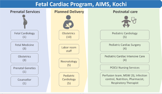
A multi-disciplinary approach is required to provide an adequate counseling and then to manage the condition through the perinatal period. All these services are provided by the Fetal cardiology Division at AIMS (see picture below).
Early scan at 14- 16 weeks
- Previous child with birth defect of heart
- Abnormal heart in routine obstetric scan
- Irregular heart beat (both fast and slow)
- Hydrops fetalis (abnormal fluid collection)
- Increased first trimester nuchal fold thickness > 2.5 mm
- Chromosomal anomaly in fetus
Routine scan at 18-20 weeks *
- Diabetes mellitus in pregnant mother
- Exposure to harmful medicines or toxins
- Intra-uterine infections
- Abnormal antibodies in mother (auto-immune disease)
- Major abnormalities of other organs detected in antenatal scan.
- Babies conceived by in-vitro fertilization (IVF) or intra-cytoplasmic sperm injection (ICSI)
Contact Us
Fetal Cardiology Division,
Department of Pediatric Cardiology
Amrita Hospital, Kochi, Kerala, India
Telephone: 0484 -2853570; Fax: 0484 – 2802020.
Email: [email protected]; [email protected]
Dedicated on-call mobile/Whatsapp: +91 7994 999 773 (for appointments)
Overview
The fetal cardiology division at Amrita Hospital, Kochi was launched in January 2008. The aim of this service is to create awareness among the medical community regarding the need and potential benefits of prenatal diagnosis of birth defects of the heart. This will provide the concerned families with more options for management rather than facing the trauma of taking a decision after the baby is born. The concept of prenatal diagnosis and planned peri-partum care for fetuses with critical heart defects was pioneered by us in India and South Asia. The division is very active academically and has been instrumental in the training of healthcare professionals in fetal heart evaluation globally and offered training courses and fellowships in fetal cardiology for doctors (both in-person and online). The division has published several important research papers in the field of Fetal Cardiology in various peer reviewed national and international journals. The division is spearheaded by Prof. Balu Vaidyanathan, who is renowned globally for his contributions to the field of Fetal Cardiology.
Doctors
Why choose us?
- Pioneered prenatal diagnosis and planned perinatal care for fetuses with critical heart defects in India.
- Provides delivery services and expedited neonatal cardiac care under one roof.
- Established in 2008, with over 10,000 fetal echocardiography studies conducted so far.
- Enabled planned delivery of more than 750 women carrying fetuses with critical heart defects at Amrita Hospital, Kochi.
- Performed over 500 neonatal cardiac procedures with a success rate of over 98%.
- Treated fetal heart rhythm disorders in 100+ fetuses through in-utero therapy.
- Performed the first successful in-utero fetal aortic valve dilatation in 2015.
What We Offer
- Comprehensive care and treatment for various heart problems in the developing fetus.
- State-of-the-art diagnostic equipment, Voluson Expert 22 and 4D STIC Ultrasound, for early detection of fetal heart issues.
- A multidisciplinary team of specialists, including fetal cardiologists, fetal medicine experts, and fetal genetics specialists, makes the treatment decisions.
- Counselors to educate and help parents in the decision-making process
- Service coordinators to ensure seamless care for a fetus with a heart defect
- Close collaboration with allied departments, including obstetrics, radiology, pediatric cardiology, pediatric cardiac surgery, and neonatology, for better outcomes
Service
- Comprehensive fetal cardiac imaging and diagnosis
- 4D STIC Fetal echocardiography and Fetal Quantification ( fetal HQ analysis)
Facilities
- A dedicated multidisciplinary team of experts to provide flawless care for a fetus with heart disorders
- Expert management and support during pregnancy, birth, neonatal, and extended post-natal care
- Most advanced technology- including Voluson Expert 22 Ultrasound equipment with its full array of transducers and fetal HQ software- for fetal heart function assessment.
- Accurate diagnosis of fetal heart defects through real-time and offline analysis of fetal heart function evaluations.
- Parental counseling to support informed decision-making.
- Highly specialized and expedited cardiac care for babies born in our center following a prenatal diagnosis.
Fellowship Programmes/Trainings
Gallery
FAQs
Fetal echocardiography is a highly specialized form of antenatal scan where a detailed evaluation of the unborn baby’s heart is carried out. This enables diagnosis of various forms of birth defects of the heart (congenital heart defects) and heart beat disorders (rhythm problems) in the fetus.
The principle of ultrasound scan is used for this technique. A minimum requirement for screening the fetal heart is a combination of 4 chamber view (heart chambers, valves and partitions), outflow tract view and 3-vessel view (blood vessels) which enables most of the cardiac defects to be detected. A detailed fetal echocardiography requires a thorough knowledge about the various types of birth defects of the heart and what all treatment can be offered including the costs involved. Few centers in India have a specialized fetal cardiology unit that provides a comprehensive facility for diagnosis, counseling and then treatment for various birth defects of the heart.
The ideal timing of fetal heart scan would be around 18-20 weeks. It is preferable to conduct these scans sufficiently early in pregnancy, so that if a problem is diagnosed, the family can be offered all possible options for management. Table 1 summarizes indications for fetal heart scan.
Fetal echocardiography uses the common ultrasound principle and it has been shown without any doubt that this is very safe, even if performed multiple times, in pregnancy.
Pre-natal diagnosis offers a much wider options to the expectant family, especially in the setting of developing countries with limited resources to treat complex heart defects.
Early diagnosis of very complex cardiac defects and multi-system disorders (cardiac defects with associated anomalies like genetic disorders) offers families the option of medical termination of pregnancy. This maybe a practical approach for very complex defects that can only be offered palliative procedures after birth with sub-optimal long-term outcomes.
Pre-natal diagnosis may possibly improve the post-natal outcomes of infants with critical but correctable heart defects (Eg: transposition of great arteries (TGA) or duct dependent conditions) by in-utero transport to a pediatric cardiac facility. Our data shows the dual benefit of improved neonatal outcomes along with cost savings for such critical defects.
Certain forms of heart defects (especially heart beat problems) can be offered in-utero treatment ( trans-placental therapy) with excellent outcomes to the fetus.
A multi-disciplinary approach is required to provide an adequate counseling and then to manage the condition through the perinatal period. All these services are provided by the Fetal cardiology Division at AIMS (see picture below).
Early scan at 14- 16 weeks
- Previous child with birth defect of heart
- Abnormal heart in routine obstetric scan
- Irregular heart beat (both fast and slow)
- Hydrops fetalis (abnormal fluid collection)
- Increased first trimester nuchal fold thickness > 2.5 mm
- Chromosomal anomaly in fetus
Routine scan at 18-20 weeks *
- Diabetes mellitus in pregnant mother
- Exposure to harmful medicines or toxins
- Intra-uterine infections
- Abnormal antibodies in mother (auto-immune disease)
- Major abnormalities of other organs detected in antenatal scan.
- Babies conceived by in-vitro fertilization (IVF) or intra-cytoplasmic sperm injection (ICSI)
Contact Us
Fetal Cardiology Division,
Department of Pediatric Cardiology
Amrita Hospital, Kochi, Kerala, India
Telephone: 0484 -2853570; Fax: 0484 – 2802020.
Email: [email protected]; [email protected]
Dedicated on-call mobile/Whatsapp: +91 7994 999 773 (for appointments)