Please wait while we prepare everything...
Please wait while we prepare everything...
Please wait while we prepare everything...
Please wait while we prepare everything...
Please wait while we prepare everything...
Please wait while we prepare everything...
Please wait while we prepare everything...
Please wait while we prepare everything...
Please wait while we prepare everything...
Please wait while we prepare everything...
Please wait while we prepare everything...
Please wait while we prepare everything...
Your health is our priority. Amrita Hospital ensures you and your family receive the best possible medical care and assistance. We strive to create a warm and safe healing environment for you and your family. Over the past decade, Amrita has been unflinchingly devoted to improving healthcare and treatment. Medical specialists have been working diligently to conduct research and educate future generations of doctors and healthcare workers.
As our entire team works toward your speedy recovery, we utilize highly-trained doctors and cutting-edge technology in the field of medical sciences.

The Comprehensive Andrology & Male Sexual Health Clinic is a specialized centre dedicated to addressing the unique health concerns of men across different stages of life. Designed as a safe and confidential space, the clinic brings together experts from multiple disciplines to evaluate and manage conditions related to male fertility, hormonal balance, and sexual well-being.

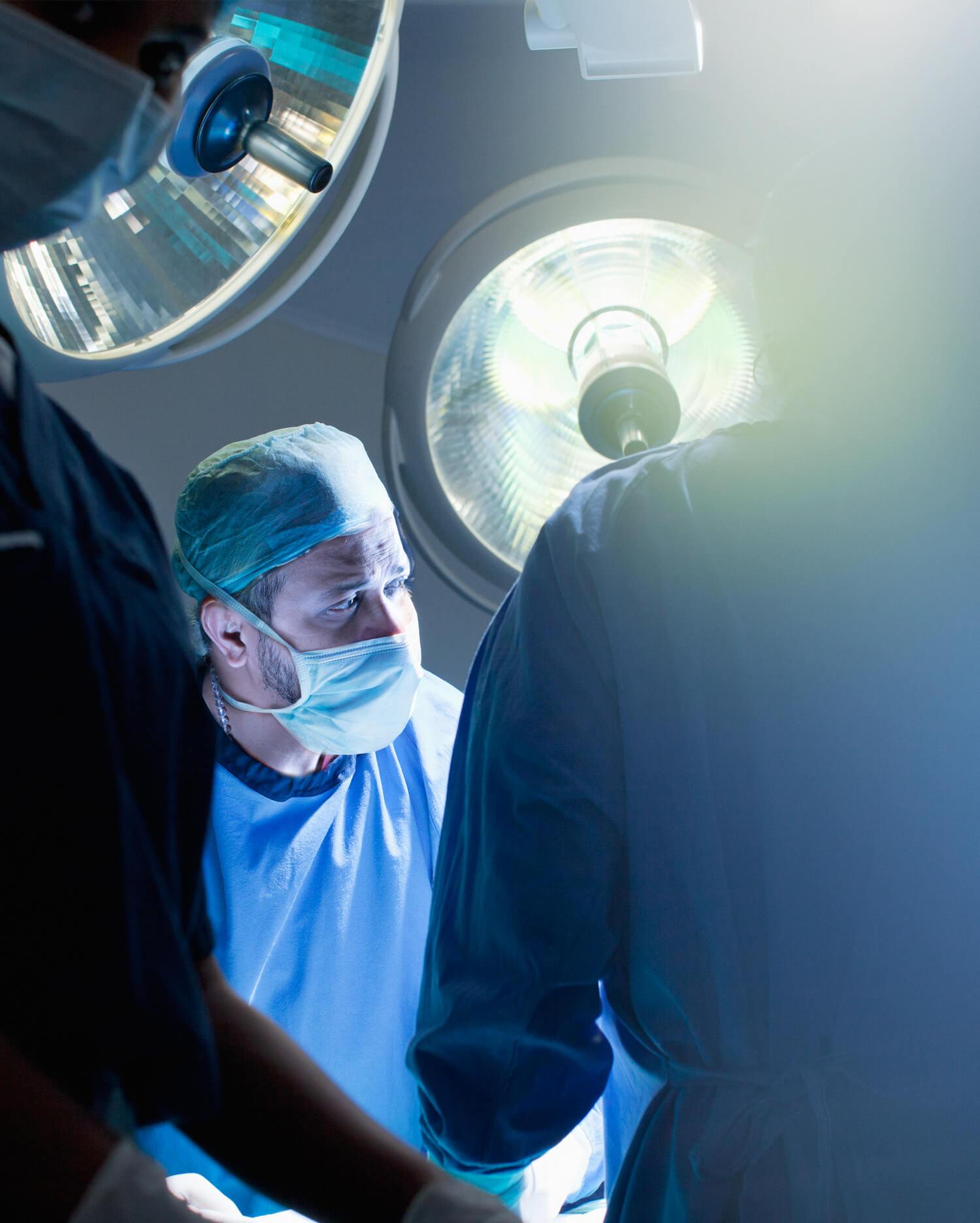
The Cardiac Anesthesia Department at Amrita Hospital specializes in providing comprehensive care for patients undergoing cardiac surgeries and procedures. Our team of highly skilled and experienced cardiac anesthesiologists is dedicated to ensuring the safety and comfort of patients throughout their cardiac interventions.

The Department of Dermatology & Cosmetology at Amrita Hospital offers procedures and services, both investigative and curative, pertaining to general dermatology, cosmetic dermatology and venereology. Comprehensive consultation and treatment is provided for both outpatients and in-patients covering all dermatological conditions including:

Amrita Hospital has been the torch bearer of this revolutionary change from the “MBBS doctor/CMO” managed casualty to ER managed by trained Emergency Medicine Physicians. With the first 3-year postgraduate training program (MD-Emergency Medicine) in Kerala, the Department of Emergency Medicine has grown into a reputed center for emergency care in India.

Our society is rapidly undergoing change. Families are becoming nuclear. The vast majority of the young are moving away in search of jobs and careers. These include women who were traditional caregivers to the elderly. We are now facing a situation where the elderly are increasing in numbers and living longer while the number of care givers is rapidly decreasing.

The Department of General Medicine at Amrita Hospital, Kochi is one of the largest general medicine department in Kerala. Our department serves as a central hub for comprehensive medical care with special emphasis on diagnosis and treatment of patients with complex medical illnesses. Also focussed on education and research to improve the patient outcome and advance medical knowledge. The department of General Medicine follows an integrated approach in inpatient and outpatient care and aims at providing seamless continuity from diagnosis to treatment and follow-up.

The Department of Paediatric Pulmonary and Critical Care at Amrita Hospital is the first Centre of Excellence in South India dedicated to the diagnosis and management of children with acute and chronic respiratory conditions and critically ill children needing organ support of various kinds.
The Department of Pathology provides comprehensive diagnostic services with emphasis on accuracy, precision and prompt reporting to our patients thereby helping and guiding the clinicians in proper patient management. The department started in 1998 along with the inauguration Amrita Hospital, and since then is located at 6th tower, 3rd floor with easy accessibility to all the clinicians.
The Poison Control Centre of Amrita Hospital, Cochin, Kerala was established in 2003. It has a Poison Information Unit that answers queries on poisons/poisoning/envenomation/drug overdose/substance abuse from doctors or hospitals across India, and is even open to the general public.

Transfusion medicine is an integral part of patient management. Department of Transfusion medicine at the Amrita Institute of medical sciences has expanded its horizons from blood collection and component preparation to apheresis, stem cell collection and advanced testing in immunohematology. In addition, it provides expertise advice to clinicians on blood product selection and management.
Urologic-oncology is a specialized section of urology for the diagnoses and treatment of urologic cancers - i.e. - kidney, bladder, prostate, testicular, adrenal and other genito-urinary cancers. The focus on cancer management enables a patient to get the best possible treatment and outcomes.

Offering the most comprehensive laboratory with the best advancements in technology.
Read more
Delivering the best possible care and treatments to patients in a morally responsible and compassionate manner.
Read more
Experience unparalleled healthcare services right at your doorstep with Amrita Home Care. Our dedicated team of professionals offers a wide range of in-home medical services, ensuring compassionate, high-quality care tailored to meet your individual needs. From doctor visits and palliative care to nursing procedures and home physiotherapy, we are committed to providing the best possible outcomes in the comfort of your home.
Read more

We collect and test your blood samples and process hematopoietic stem cells with the utmost care and in a very responsible manner.
Read more
Amrita Hospital is the first university teaching hospital to get NABH accreditation in the country. The National Accreditation Board for Hospitals & Healthcare Providers (NABH) is a constituent board of Quality Council of India, set up to establish and operate an accreditation programme for healthcare organizations. NABH accreditation is an assurance that healthcare providers meet specific standards of patient safety and care. It is widely recognized as a symbol of quality in the healthcare industry and can foster credibility and trust between healthcare organizations, patients, healthcare professionals, and other stakeholders.
The ISO standards provide a guarantee of quality across boundaries and geographies. Amrita Hospital in Kochi is accredited with ISO 9001:2015 standards. ISO 9001:2015 is a standard for quality management systems that emphasizes customer satisfaction, continuous improvement, and regulatory compliance. By following these requirements, organizations can strive to improve the safety and quality of care they provide, and consistently meet the expectations of both customers and regulatory bodies.
The National Accreditation Board for Testing and Calibration Laboratories (NABL) is a board that operates under the Quality Council of India. It is responsible for providing accreditation to testing and calibration laboratories in India, and for promoting the quality of testing and calibration services in the country. By achieving NABL accreditation, our laboratory has demonstrated its ability to perform accurate and reliable tests and measurements, and to ensure that its results are traceable to national or international standards. NABL accreditation also indicates that the laboratory has implemented a quality management system that adheres to international standards and best practices.

The Stroke Medicine Department at Amrita Hospital has been officially assessed and certified by the National Accreditation Board for Hospitals & Healthcare Providers (NABH) and the World Stroke Organisation (WSO) as an Advanced Stroke Centre.


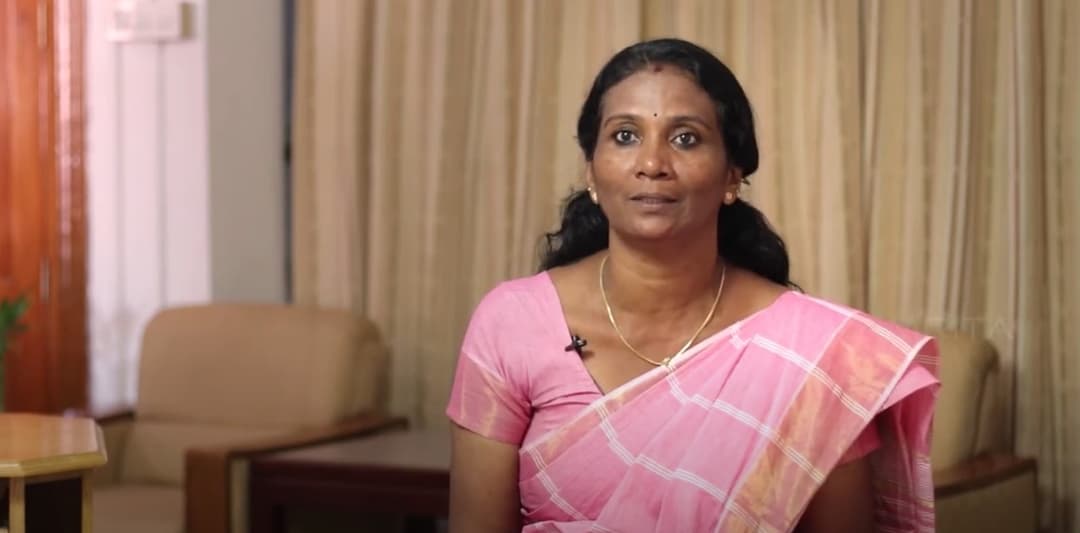
Connect to our expert doctors, who share their insights and knowledge to help you heal and recover. From practical tips to inspiring stories, we offer a wealth of information to help you heal faster and get back to a healthy, happy life. Join us on our journey to better health.