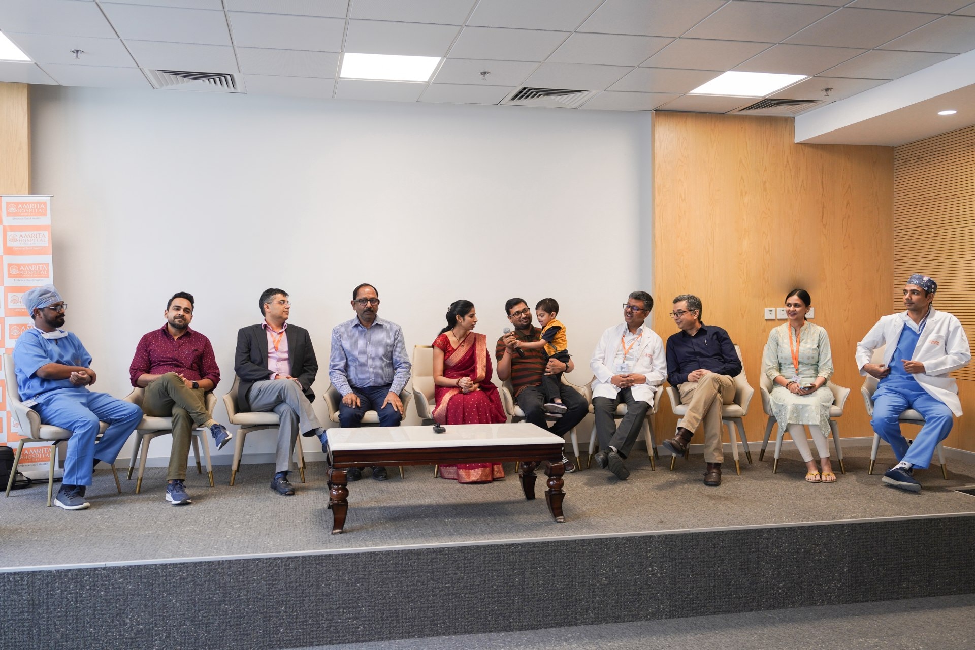
A four-year-old boy born with Median Facial Cleft Syndrome, a very rare facial deformity that bisects (divides) the face vertically through the midline due to improper fusion of bone and tissue, was successfully operated upon by doctors at Amrita Hospital, Faridabad. The marathon surgery, that lasted 18 hours, restoring the young child’s appearance, giving him a new lease of life.
The boy, whose father works in Gurugram in a multinational IT firm, was suffering from a midline gap in the face that existed on the skin and hard tissue, extending up to the skull base and brain. The gap caused an increased distance between both his eyes (Orbital Hypertelorism). There was also cleft of lip, palate, and nose, and Basal Encephalocele (a pouch of brain tissue herniating from the skull base into the nasal cavity). These anomalies led to problems related to binocular vision, speech and feeding, and psychosocial and aesthetic challenges. The parents were home schooling the child due to worries that he would be teased at school or other kids would get scared seeing his face.
Said Dr. Mohit Sharma, Head, Department of Plastic and Reconstructive Surgery, Amrita Hospital, Faridabad: "We have the most comprehensive department of plastic and reconstructive surgery, which can cater to all Craniofacial anomalies. We have state-of-the-art facilities for carrying out complicated surgeries that last for hours. The surgery was done by a team of 10 doctors," adding that the surgeons rehearsed for the surgery virtually as well as physically through mock surgeries on 3D printed models. The mock and virtual surgery enabled them to visualise the extent of corrective measures to be made.
The complex surgery was led by renowned Craniofacial Surgeon, Dr. Anil Murarka, Senior Consultant, Department of Plastic and Reconstructive Surgery, Amrita Hospital, Faridabad along with Dr. Arun Kumar Sharma, Consultant (Cranio-Maxillofacial Division), Amrita Hospital, Faridabad.
Said Dr. Murarka and Dr. Sharma: "Midline facial clefts are extremely rare, occurring in about 1 in a million live births. The boy is a special child, as not only was his condition very rare, the deformity was also extremely severe. Orbital hypertelorism (abnormal distance between the eyes) is graded as type I, II, and III, with Type III being the most severe at more than 40mm. The boy had more than 60mm distance between the eyes. The corrective surgery in such cases is called facial bipartition and is performed by craniofacial surgeons."
They added: "We focused on correcting the boy’s facial cleft (cleft of lip and nose) and reducing the distance between the eyes. The two halves of his face were brought together in the midline after removing excess bone and soft tissue between the eyes. He also required a separate surgery for the removal of herniating brain and reconstruction of the skull base defect. This was performed by a team of pediatric neurosurgeons."
Said Dr. Anurag Sharma, Consultant, and Dr. Sachin Gupta, Consultant, Department of Paediatric Neurosurgery, Amrita Hospital, Faridabad: “It was an extremely complex surgery which required teamwork from various specialties like neuro-anesthetists, pediatric neurosurgeons and craniofacial surgeons. In view of the severity of the case, a lot of hard work and planning was done by the surgeons to make sure of every step. They rehearsed for the surgery virtually as well as physically through mock surgeries on 3D printed models. This enabled us to visualize the extent of correction required. Blood auto-transfusion machine and multiple units of blood and blood components were also arranged. However, no preparation was enough until the surgery got actually performed on the vulnerable child, and even the more delicate brain and eyes.”
The patient was shifted to pediatric ICU on ventilatory support following the surgery. For the first 2-3 days, his vitals were stable and he was breathing well with his own efforts. However, he was not completely arousable. Post-operative CT scans showed a basal infarct (tissue death due to lack of blood supply) in a part of the brain. The doctors also noticed slight hemiparesis (inability to move) of the right side of the body.
Said Dr. Maninder Singh Dhaliwal, Senior Consultant, and Dr. Veena Raghunathan, Senior Consultant of the Department Pediatric Critical Care, Emergency & Respiratory Medicine: "The long surgery had taken its toll on the delicate brain of child. Our pediatric intensive care team decided to tracheostomise the patient (a procedure to help air reach the lungs by creating an opening into the trachea or windpipe from outside the neck). This helped the doctors take the boy off the ventilator. Now, we were waiting for him to recover neurologically. The patient spent three weeks at the hospital, after which he was discharged. After a month and half, he is now doing well. The boy is using both his hands well and walking on his own. It would probably take 3-4 more weeks to come back to his usual playful self. We now hope to see him go to school soon and resume his daily social life with pride and confidence."
The anesthesia team led by Dr. Gaurav Kakkar, Senior Consultant, along with Dr. Ketan Kulkarni, Senior Consultant, and Dr. J. S. Rahul, Consultant from the Department of Anesthesiology, Amrita Hospital, Faridabad played a major role in conducting the 18-hour surgery.
Said the boy’s father: "My son's condition is improving every day, and we are all so excited at see him recover and go to school like any other child of his age. I thank the doctors of Amrita Hospital from the bottom of my heart at making this extremely complex surgery a success and giving my child a second chance at life."


